CRFiCOR: Imaging Core Labs
Services
CRF Imaging Core Labs: Imaging Experience, Leadership and Vision
CRF Imaging Core Labs is the name for the CTC’s comprehensive cardiovascular imaging laboratories, which are directed by talented and experienced physician leaders who specialize in all aspects of coronary angiography, carotid and peripheral angiography, intravascular imaging, and the assessment of structural heart disease. The Angiographic Core Lab and Intravascular Imaging Core Lab have consistently advanced the field by developing and validating new methodologies.
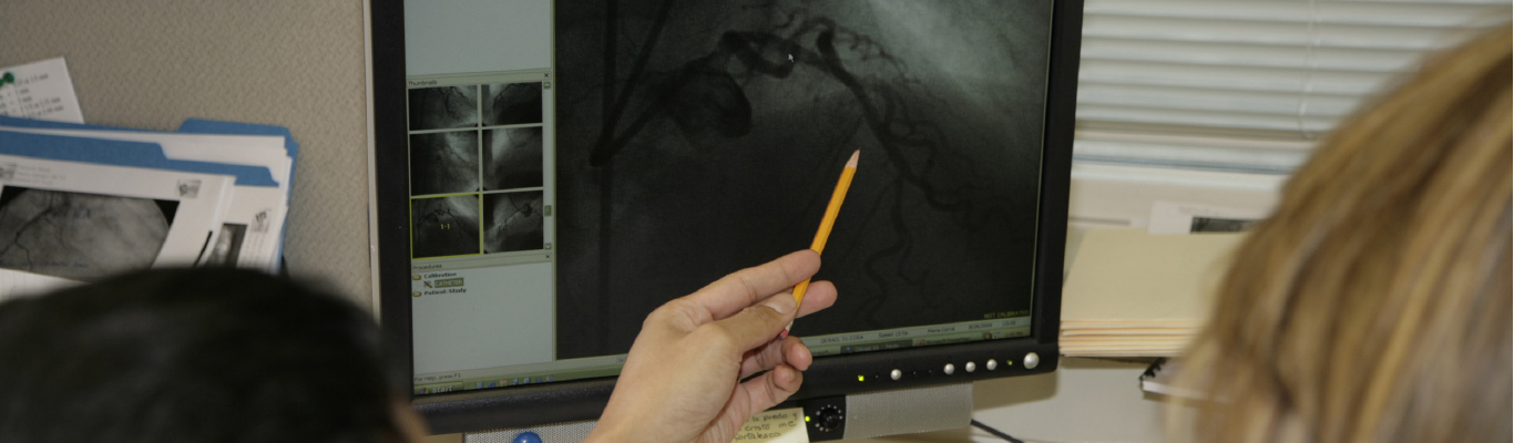
Angiography (Coronary, Peripheral, Valve, and Structural Heart Disease)
Led by an experienced and dedicated team, the Angiographic Core Lab (ACL) has over 20 years of coronary, peripheral, structural, and physiological angiogram analysis experience with an annual volume of over 20,000 cases.
Capacity and Commitment
The ACL currently employs highly trained, experienced analysts dedicated to performing qualitative and quantitative assessment for coronary and peripheral artery therapeutics. We have extensive experience in trials for BMS, DES, BVS, and DCB, as well as other novel devices or drugs, and will tailor the angiographic case report form to collect robust and meaningful data based on the study protocol. All analyses are 100% over-read as a quality measure to ensure completeness and accuracy. We have the capability to process a high volume of cases while maintaining excellent quality and reproducibility.
5-Day Turnaround Time
- We recognize the importance of providing timely results to our clients. Our Angiographic Core Lab is accustomed to working on high-volume cases while ensuring that time requirements are met.
Accomplishments
- Analysis of more than 100,000 patients in more than 550 trials
- Experienced in first-in-human, pre-market approval, and post-marketing surveillance
- More than 50 Investigational Device Exemption (IDE) trials
Echocardiography
There are few non-invasive and non-radiographic imaging tests that can yield as much information about the heart as the echocardiogram. The Echo Core Lab reviews and develops all echocardiographic imaging protocols, policies, and procedures. In addition, site selection and training are an important component of our Core Lab responsibilities. Our experience and capabilities include:
- Two-dimensional and Doppler echocardiography
- Quantitative assessment of ventricular structure and function
- Quantitative assessment of valvular structure and function
- Three-dimensional echocardiography
- Structural and functional assessment
- Doppler assessment
- Tissue Doppler and Speckle Strain imaging
Electrophysiology, Cardiac Rhythm Management and ECG
Electrocardiography and Rhythm Core Lab
The Electrocardiography and Rhythm Core Lab supports large-scale national and international trials by providing independent electrocardiographic interpretation. Our readers are board-certified academic electrophysiologists with expertise in analysis of all types of electrocardiographic data. The services provided include:
- Electrocardiographic, ICD and pacemaker rhythm analysis; remote monitoring, utilization, and interrelationship with other outcomes
- Rhythm monitoring for Holter monitors, event recorders, and for implantable devices including loop recorders, pacemakers and defibrillators
- Analysis of ECG intervals, including QT and corrected QT intervals, morphology, conduction and rhythm abnormalities, and expert evaluation of ST-segment deviation
- ST segment elevation and resolution data, a key component of interventional efficacy in many trials, can be customized for your study
As leaders in the industry, the CTC staff will work with you to define and fulfill the ECG and rhythm analysis needs for your study.
Hemodynamics
An increasing number of heart failure studies involve the collection and interpretation of hemodynamic data. Clinically used hemodynamic recording systems (in a cath lab or intensive care unit) generally include software that provides automated readings of all common hemodynamic parameters. While physicians generally rely on these readings for clinical decision making, these algorithms lack certain sophistication required for detailed analyses as required in the context of clinical trials. Furthermore, different recording systems employ different algorithms for quantifying recordings. This is particularly the case when hemodynamics are being recording during exercise, which is being included in a growing number of studies. To overcome these limitations and to overcome the potential for introduction of investigator bias a Hemodynamic Core Lab is recommended to ensure consistent, systematic collection and interpretation of data.
Specific services include:
- Generation of study-specific SOPs for performance of hemodynamic measurements including:
- Equipment setup
- Calibration
- Recordings
- Site training on the SOPs and validation
- Development of study-specific worksheet for use by the sites during testing
- Logging and tracking of tests received
- Rapid feedback on test quality
- Provide retraining if necessary
- Blinded interpretation of study results
- Study-specific hemodynamic results database
- Statistical analysis of study results, expert interpretation of findings and report generation
Intravascular Imaging (IVUS, VH-IVUS, NIRS, and OCT)
The Intravascular Imaging Core Lab has been a pioneer in the intravascular imaging field. In conjunction with our core group of researchers, our exceptional team of international cardiology fellows focuses on academic pursuits such as developing abstracts, presentations, and publications. Our core expertise includes the following intravascular modalities:
- Grayscale Intravascular Ultrasound (IVUS)
- Virtual Histology IVUS (VH-IVUS)
- Optical Coherence Tomography (OCT)
- Near-Infrared Spectroscopy (NIRS)
- Fractional Flow Reserve (FFR)
Our expertise includes:
- Innovative and adaptive intravascular imaging study structure and design
- In-house programming for analysis of novel imaging technologies
- Industry leadership in the formulation of IVI standards and definitions
- Pioneering some of the largest IVUS studies to date
- Seminal contributions to the field of intravascular ultrasound
Our Core Lab has been actively contributing to the field of intravascular imaging for more than two decades, developing a wealth of knowledge and experience in the field.
Accomplishments
- Intravascular ultrasound (IVUS), virtual histology-IVUS, optical coherence tomography (OCT), and near-infrared spectroscopy (NIRS)
- Analysis of more than 50,000 cases and publication of more than 400 papers
- Training site for more than 50 fellows from around the world
Non-Invasive Imaging (CT, MRI)
The Cardiac Computed Tomography Angiography (CCTA) Core Lab performs comprehensive qualitative and quantitative analyses on cardiovascular computed tomographic angiograms. The CCTA Core Lab provides coronary artery analysis for volumetric quantification and characterization of atherosclerotic plaque and vessel measures; cardiac analysis for quantitation of myocardial function and perfusion; valvular function and measurements; and other vascular evaluations.
State-of-the-art advances in imaging technology now permit non-invasive evaluation of coronary, cardiac, and vascular findings with very high accuracy compared to invasive imaging. The CCTA Core Lab also provides assistance with developing CCTA protocols, image-acquisition methods, site qualification, image feedback and training.
MRI
The Cardiac Magnetic Resonance Imaging (MRI) Core Lab assesses global and regional systolic ventricular function, myocardial perfusion and viability, and valvular heart disease. Recent advances in the field have dramatically improved the quality of cardiac MRI images by providing:
- Excellent spatial and temporal resolution
- Higher precision and reproducibility
- Adequate sensitivity to detect smaller areas of myocardial damage with an accuracy surpassing other forms of testing, allowing trials to reach statistical significance with fewer patients.
- Assesses global and regional systolic ventricular function, myocardial perfusion and viability and valvular heart disease
- Provides qualitative and quantitative analyses focusing on infarct size and residual myocardial viability
Cardiovascular Physiology
The Cardiovascular Physiology Core Lab at the Cardiovascular Research Foundation provides researchers, clinicians, and clinical trial sponsors with state-of-the-art centralized analysis of cardiac and coronary physiology data. Coronary physiology measurements are increasingly used clinically as adjunctive tools during diagnostic and interventional coronary procedures. As the body of research expands, further establishing the value of these modalities, quality control of future clinical studies will become increasingly important. Accordingly, the mission of the Cardiovascular Physiology Core Lab is to provide a novel service of ensuring data quality for clinical research trials, which heretofore has not been standard practice in coronary physiology studies.
Services are tailored to the investigator’s needs and focus on providing data suitable for use in future studies, FDA submissions, and scientific manuscripts. Analyses provided include objective waveform assessment to ensure adequate data quality and independent calculation of the various physiology indexes.
Our comprehensive services include:
- Analysis of Fractional Flow Reserve (FFR) measurements
- Analysis of Coronary Flow Reserve (CFR) measurements
- Assessment of various indexes of microvascular function (e.g. , index of microvascular resistance [IMR])
- Evaluation of various physiology resting and exercise indexes